A) MRI findings: the typical bunched medial collateral ligament (MCL)

By A Mystery Man Writer
Download scientific diagram | (A) MRI findings: the typical bunched medial collateral ligament (MCL) fibres are obvious on the T2-weighted MR image (arrow). Countercoup oedema is evident in the lateral tibial plateau. (B) Anatomical findings: the fibres are short and abruptly jump over the semitendinosus tendon. The femoral insertion site remained intact. Note. sMCL, superficial MCL. from publication: Isolated medial collateral ligament tears: An update on management | Tears of the medial collateral ligament (MCL) are the most common knee ligament injury. Incomplete tears (grade I, II) and isolated tears (grade III) of the MCL without valgus instability can be treated without surgery, with early functional rehabilitation. Failure of | Tears, Collateral Ligaments and Reconstruction | ResearchGate, the professional network for scientists.

A–B MRI findings of the left knee of the first patient are shown

MRI description of knee medial collateral ligament abnormalities
The Do's and Don'ts After ACL & MCL Tears & Surgery

A) MRI findings: the typical bunched medial collateral ligament (MCL)

Subgroup analysis for IKDC score at follow-up according to

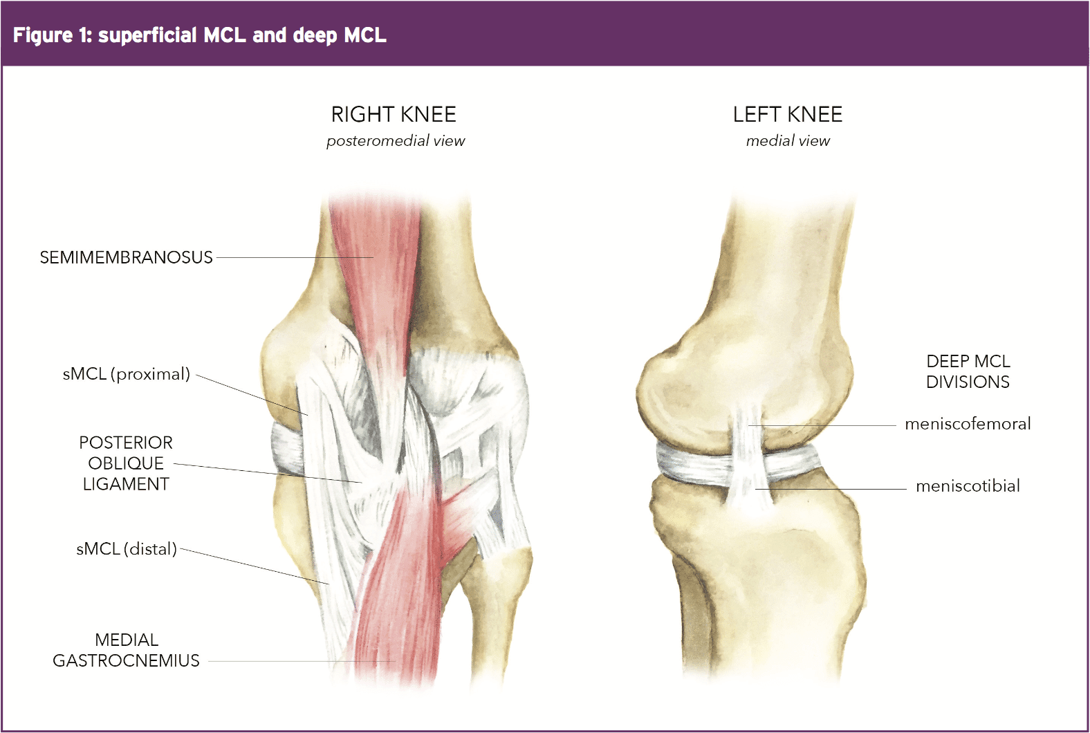
High Prevalence of Superficial and Deep Medial Collateral Ligament

Single-Row Repair in Chronic Medial Collateral Ligament

Isolated medial collateral ligament tears: An update on management. - Abstract - Europe PMC
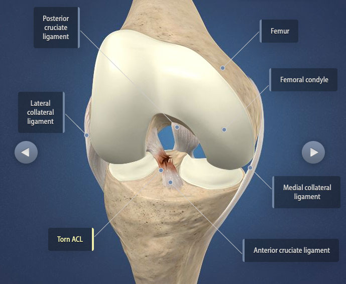
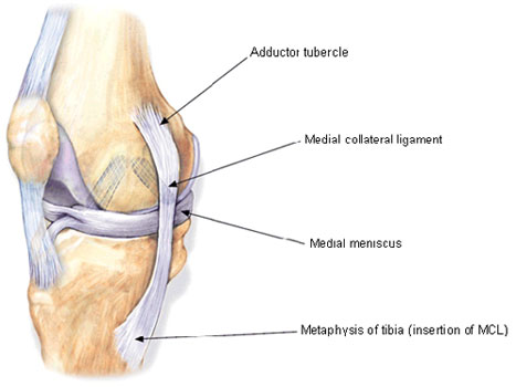
MCL: Anatomy, Biomechanics & Injury Science

- Hanes Sport Men’s Air Mesh Long Leg Boxer Brief Underwear, X-Temp, 4-Pack Assorted XL

- See How She Got Started: OUA & McMaster University Tennis Player Maia Mureseanu - SWSCD

- Women's Pajama Pants Stretch Pant Drawstring Lounge Pants, Watercolour Art Floral Flower Spring Yellow Tulip : : Clothing, Shoes & Accessories

- Selecting appropriate compression for lymphedema patients

- Dx Point BLACK LYCRA STRETCHABLE PETTICOAT Lycra Blend Petticoat Price in India - Buy Dx Point BLACK LYCRA STRETCHABLE PETTICOAT Lycra Blend Petticoat online at