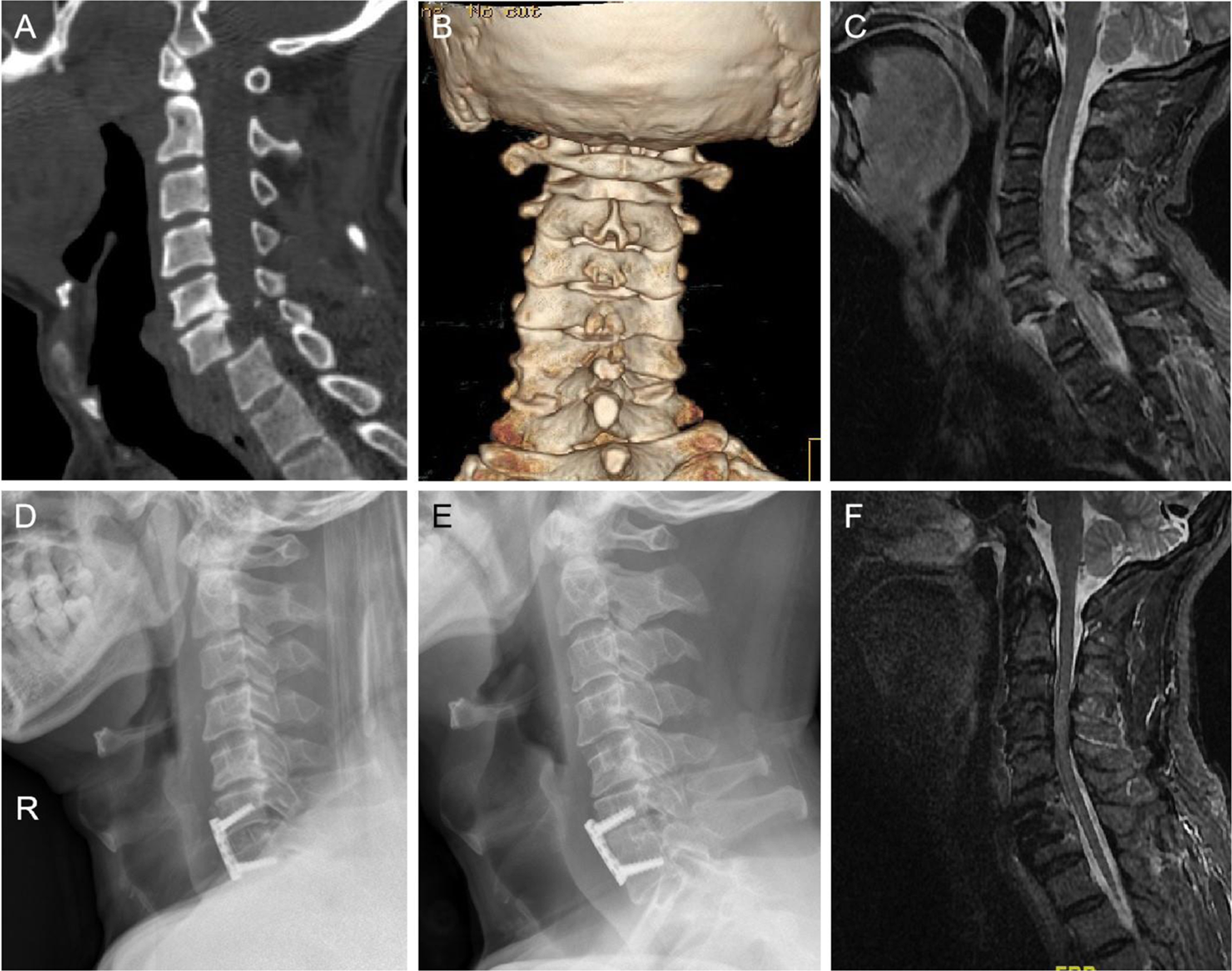
Lateral cervical spine showing C0-C3 fusion in reduced position

By A Mystery Man Writer

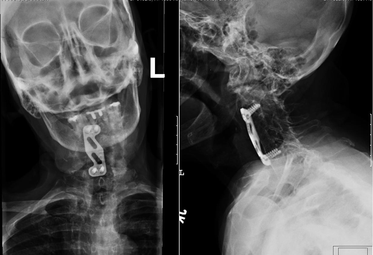
Intraoperative C-spine lateral (A) radiograph using the
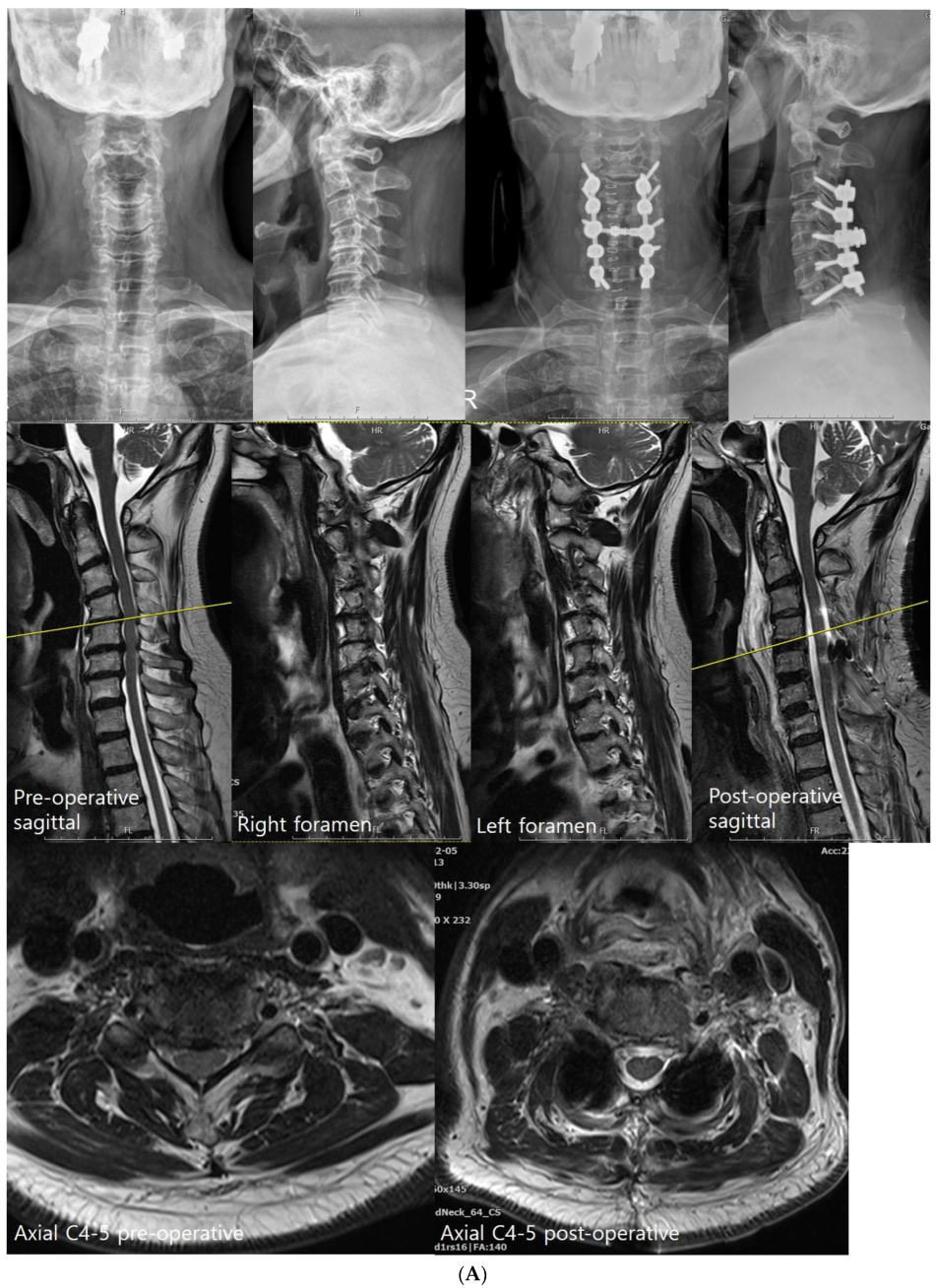
Posterior Cervical Fusion Excluding the Occiput
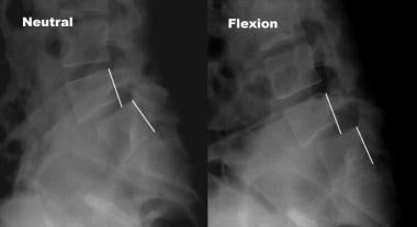
Lateral dynamic views of the cervical spine in flexion (A) and

Correlation between postoperative C1-C2 and C2-C7 angle. Postoperative

Rui PINTO, Clinical Director, MD, Head of Department of St.Maria- Porto Hospital, Orthopedics and Traumatology

Nuno ALEGRETE, Research Assistant, University of Porto, Porto, UP, Instituto de Engenharia Biomédica (INEB)

Symptoms and conditions of Craniocervical and Cervical Instability – Caring Medical Florida
JCM, Free Full-Text

Lateral cervical spine showing C0-C3 fusion in reduced position after

Representative load–displacement curve. Pedicle bone
Comparison of anterior and posterior approaches for treatment of traumatic cervical dislocation combined with spinal cord injury: Minimum 10-year follow-up
Spinal Instability and Spinal Fusion Surgery Workup: Laboratory Studies, Imaging Studies, Other Tests

Correlation between postoperative C1-C2 and C2-C7 angle. Postoperative
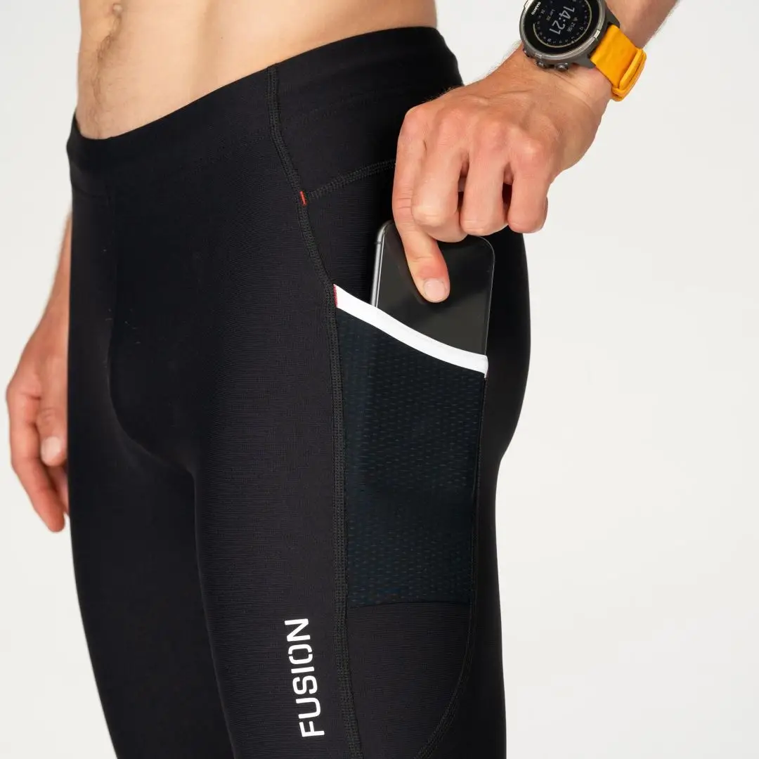
- Fusion C3 Shorts Product Review - The Best Shorts You'll Ever Own - Clean Coach Katie

- FUSION
- Fusion Produkte » Preise vergleichen und Angebote sehen
- adidas Performance TERREX SKYCHASER MID GORE-TEX® HIKING 2.0 - Hiking shoes - grey six/grey four/halo silver/grey

- Running pants: Fusion C3 Long Tights Runningtrousers - XL

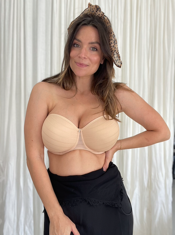
- The Best Strapless Bras For Fuller Busts! – MARVELL LANE
- S1183, Simplicity Sewing Pattern Misses' & Plus Size Corsets

- Silver Strong No Fade 304 Stainless Steel Open Split Jump Rings Connector Loop
- How to Qualify to Get On The NY Times Bestseller List
- Compression testing machine Compressive strength, Compression
