This scanning electron micrograph (SEM) depicted a number of red

By A Mystery Man Writer
Download this stock image: This scanning electron micrograph (SEM) depicted a number of red blood cells found enmeshed in a fibrinous matrix on the luminal surface of an indwelling vascular catheter; Magnified 11432x Note the biconcave cytomorphologic shape of each erythrocyte, which increases the surface area of these hemoglobin-filled cells, thereby, promoting a greater degree of gas exchange, which is their primary function in an in vivo setting. In their adult phase, these cells possess no nucleus. What appears to be irregularly-shaped chunks of debris, are actually fibrin clumps, which when inside the living organi - 2BE0H0B from Alamy's library of millions of high resolution stock photos, illustrations and vectors.

This Scanning Electron Micrograph Sem Depicted A Number Of Red Blood News Photo - Getty Images
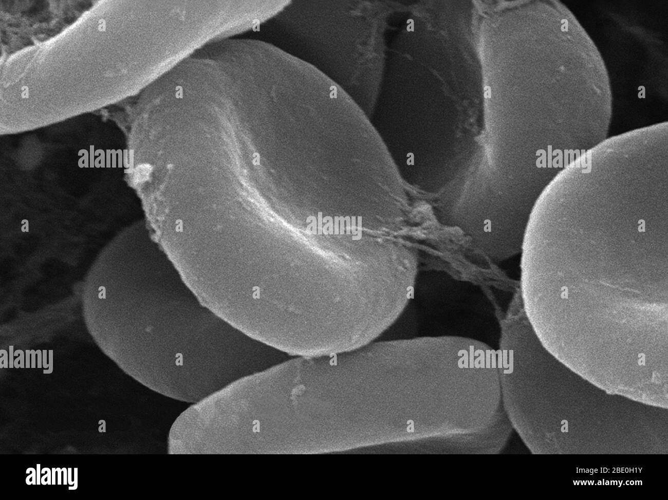
This highly enlarged scanning electron micrograph (SEM) depicted a closer look at the details exhibited by of number of red blood cells found enmeshed in a fibrinous matrix on the luminal surface

Scanning electron microscopy hi-res stock photography and images - Page 6 - Alamy
Scanning electron microscope - Wikipedia

Red Blood Cells, Sem #38 Sticker by Science Source - Pixels
165 Blood Actually Photos & High Res Pictures - Getty Images

This scanning electron micrograph SEM revealed some of the

Red Blood Cells, Sem #40 Framed Print by Science Source - Fine Art

Red Blood Cells And Acanthocyte, Sem #3 Photograph by Science
Tuberculosis Photos

Red And White Blood Cells, Sem #3 Photograph by Science Source
Scanning electron microscopy (SEM) and transmission electron
- Laboratory Evaluation of Sickle Cell Disease in the ED — Taming
- Solved Learning Guide item #7 - Describe the 3-D shape
- Hemp, Description, Products, Seeds, Fiber, & Uses

- HEMO Shapewear Women's Tummy Control Shapewear Long Pant Trainer Shaper Sexy Butt Lifter Panties Flat Abdominal Leg Shapes for Women Plus Size Corsage (Colour: Nude, Size: Large) : : Fashion

- Curved Rubber Hemo Split Bard Permanent Catheter, Size: Medium at Rs 11500 in Navi Mumbai


- Men underpants slips briefs underwear softy lingeries

- Buy Underwear for toddlers – Popup Kids
- Forever 21 Active Now Bike Shorts Womens workout outfits, Summer

- Air Ultimate Lift Bra Stretch Full-Figure Seamless Lace Cut-Out Bra,Exsecret Ultimate Lift Sports Bra,Sleep Bras for Women (48/110D, Skin Color)

- S12 Wearable Breast Pump Hands Free, MFINE Electric Breast Pump Smart Display Tire Lait Portable Breast Pumps, 2 Mode & 9 Level, Come with 24mm、21mm
