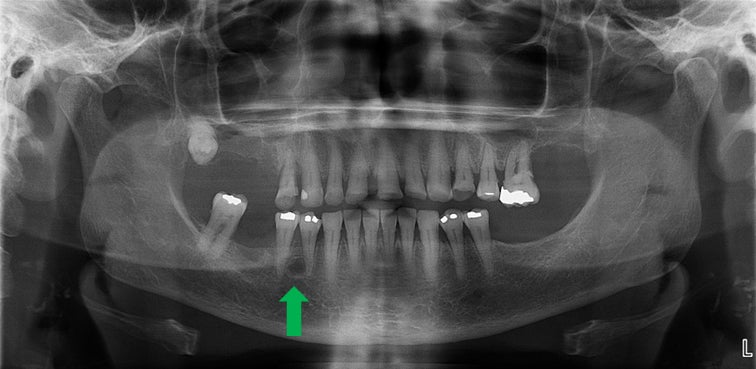
a Mandibular fistula indicated by an arrow in the apical region of dd

By A Mystery Man Writer
Download scientific diagram | a Mandibular fistula indicated by an arrow in the apical region of dd 36-37. b A fistula in the apical region of dd 46-47 (white arrows) and a red area in the mucosa (black arrows) are seen in the right lingual surface of the mandible. c Panoramic radiograph showing no bone lesions in the mandible. d Periapical x-ray with no bone involvement in the apical region of dd 46-47 from publication: Treatment of bisphosphonate-induced osteonecrosis of the jaws with Nd:YAG laser biostimulation | Osteonecrosis, Jaw and Nd:YAG Laser | ResearchGate, the professional network for scientists.

Satu ALALUUSUA, University of Helsinki, Helsinki, HY, Institute of Dentistry

JaypeeDigital

Oral Cutaneous Fistulas: Practice Essentials, Pathophysiology

Malformations of the tooth root in humans. - Abstract - Europe PMC

Diagnosis and management of benign fibro‐osseous lesions of the jaws: a current review for the dental clinician - Mainville - 2017 - Oral Diseases - Wiley Online Library

Medication-related osteonecrosis of the jaw without osteolysis on computed tomography: a retrospective and observational study

Frs hfn

JaypeeDigital

a Mandibular fistula indicated by an arrow in the apical region of dd

SciELO - Brazil - Differential diagnosis and clinical management of periapical radiopaque/hyperdense jaw lesions Differential diagnosis and clinical management of periapical radiopaque/hyperdense jaw lesions

Case Archive, School of Dental Medicine
- DD36HC-1-B, 36 Direct Draw Dispenser in Black

- Presidential bracelet question for DD 36 : r/rolex

- Tennis News 2024: Alex de MInaur beats Carlos Alcaraz amid giant killing run before Australian Open
- PAGANI DESIGN 2023 New DD36 Automatic Watch For Men Mechanical Wristwatch AR Sapphire glass stainless steel 10ATM Men's Watches

- PAGANI DESIGN DD36 Men's Watches Luxury Automatic Watch Men AR Sapphire Glass Mechanical Wristwatch Men 10Bar ST16 Movt 2023 New

- Yung Russia for Future Shift // adidas Originals x Boiler Room on
- Natori, Intimates & Sleepwear, New Natori Recharge Underwire Sports Bra

- CHICTRY Womens High Cut Backless Thong Leotard Bodysuit Nightwear Bathing Suit Beachwear

- Puff N Save - Vacationland Books

- Rockwear Evolve Moulded Adjustable High Impact Sports Bra In Sea

