Ultra-wide-field fundus photographs and ultra-wide-field

By A Mystery Man Writer
Download scientific diagram | Ultra-wide-field fundus photographs and ultra-wide-field fluorescein angiographic imaging of ocular toxocariasis. (A) A granuloma with mild vitreous opacity. (B) A tractional retinal fold with localized tractional retinal detachment. (C) Diffuse peripheral vascular leakage. (D) A prominent optic disc leakage. from publication: The Clinical Characteristics of Ocular Toxocariasis in Jeju Island Using Ultra-wide-field Fundus Photography | Toxocariasis, Ocular and Photography | ResearchGate, the professional network for scientists.
Wide-field Imaging of Retinal Diseases - touchOPHTHALMOLOGY

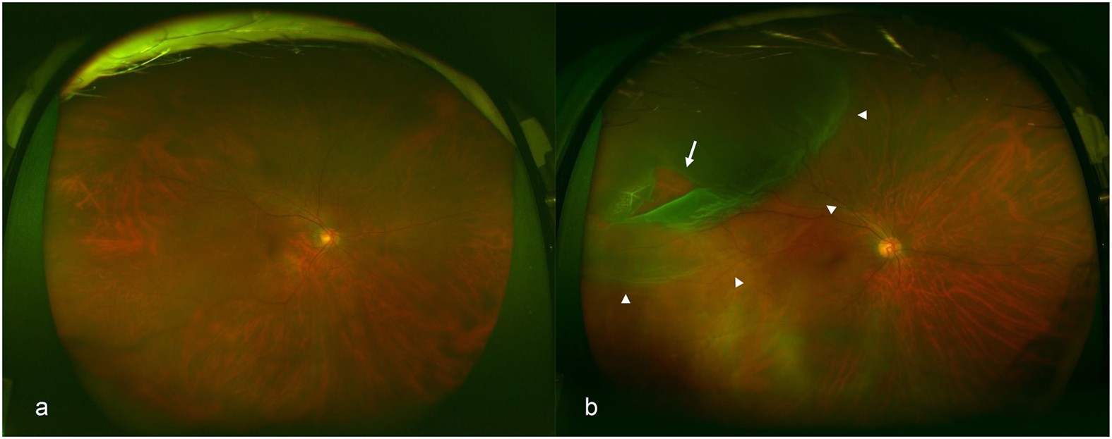
Left eye ultra-wide-field imaging covers approximately 80% of the
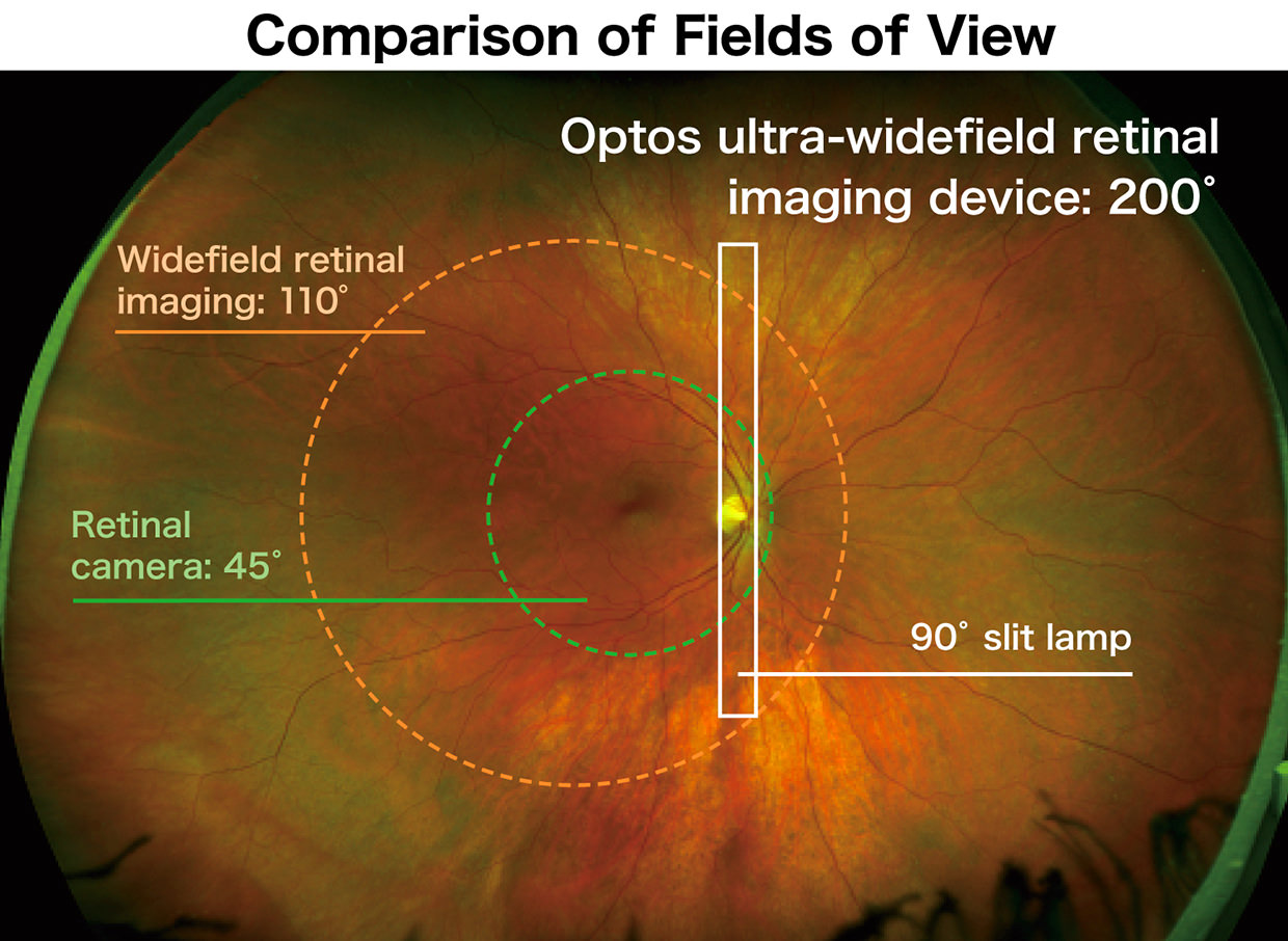
A Clearer Picture of Retinal Imaging

Sang-Yoon Lee's research works Gachon University, Seongnam-si (kyungwon) and other places
Ultra-Wide Field Retinal Imaging Device, Product Technology
Accuracy of deep learning, a machine-learning technology, using ultra–wide-field fundus ophthalmoscopy for detecting rhegmatogenous retinal detachment

Assessment of early diabetic retinopathy severity using ultra-widefield Clarus versus conventional five-field and ultra-widefield Optos fundus imaging
How Ultra-Widefield (UWF) Imaging Helps Clinically

Eun Kyoung Lee's research works Dongguk University, Seoul and other places