A 38-year-old female with increasing right breast lump since 15

By A Mystery Man Writer
Download scientific diagram | A 38-year-old female with increasing right breast lump since 15 months. Mammogram ( ): An irregular high-density mass with indistinct margins is seen in predominantly upper inner quadrant also extending in the outer quadrant measuring approximately 4.4 × 4.4 × 5.5 cm. Pleomorphic microcalcifications ( ) are seen within the mass, better seen on magnification view. Diffuse trabecular thickening with nipple areolar complex thickening and retraction is seen. Few suspicious right axillary nodes are seen, largest measuring 1.2 × 0.7 cm with 4.5-mm cortical thickness ( ). In view of dense breast parenchyma, further evaluation with CEM was performed to rule out any other lesion in breast, CEM ( ) is suggestive of large unifocal lesion. This is the case of locally advanced breast cancer (stage IIIA), further metastatic work-up was performed. On CT scan, ( ) heterogeneously enhancing mass is seen involving right breast with involvement of overlying skin. Enlarged right axillary, right internal mammary, and right supraclavicular lymph nodes are seen. (CEM, contrast-enhanced mammogram.) from publication: Imaging Recommendations for Diagnosis, Staging, and Management of Breast Cancer | In a rapidly evolving world, with a steep rise in breast cancer incidence, there has been many advances in imaging and therapeutic options of breast cancer care. In this review article, we are trying to cover imaging guideline for cancer detection and their therapeutic | Breast Cancer | ResearchGate, the professional network for scientists.

A 48-year-old woman complains of a left breast lump. a Craniocaudal and
29-Year-Old With Breast Lump Was Denied Mammogram, Has Stage 4 Cancer

Breast Lump During Pregnancy: Causes, Types of Lumps & What to Do

A Breast Mass in a 5-Month-Old Girl

Nita NAIR, Tata Memorial Centre, Mumbai, TMC, Surgical Oncology
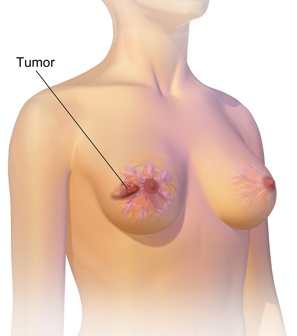
Breast Lump: When to Worry
Breast cancer - Wikipedia
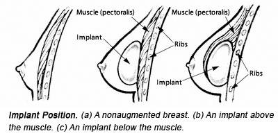
Breast Implant Placement: Over The Muscle vs. Under The Muscle

Enlarging Breast Mass in 69-Year-Old Woman With History of Cysts- Clinical Advisor

Sneha SHAH, Tata Memorial Centre, Mumbai, TMC, Nuclear medicine and Molecular imaging

Is This Normal? A Teen Guide to Breast Health

PDF) Imaging Recommendations for Diagnosis, Staging, and Management of Breast Cancer

Breast development: Stages and how to spot growth signs - Flo

Rima Pathak's research works Tata Memorial Centre, Mumbai (TMC) and other places

Complex Cystic Breast Masses: An Ultrasound Imaging Review
- Buy Generic Silicone Pocket Bra Mastectomy padded Wire Free Bra

- female between 33 and 38 years old, Brazilian, curly hair, blue

- My giant boobs saved my life! Woman claims 38KKK implants protected her in horror smash - World News - Mirror Online
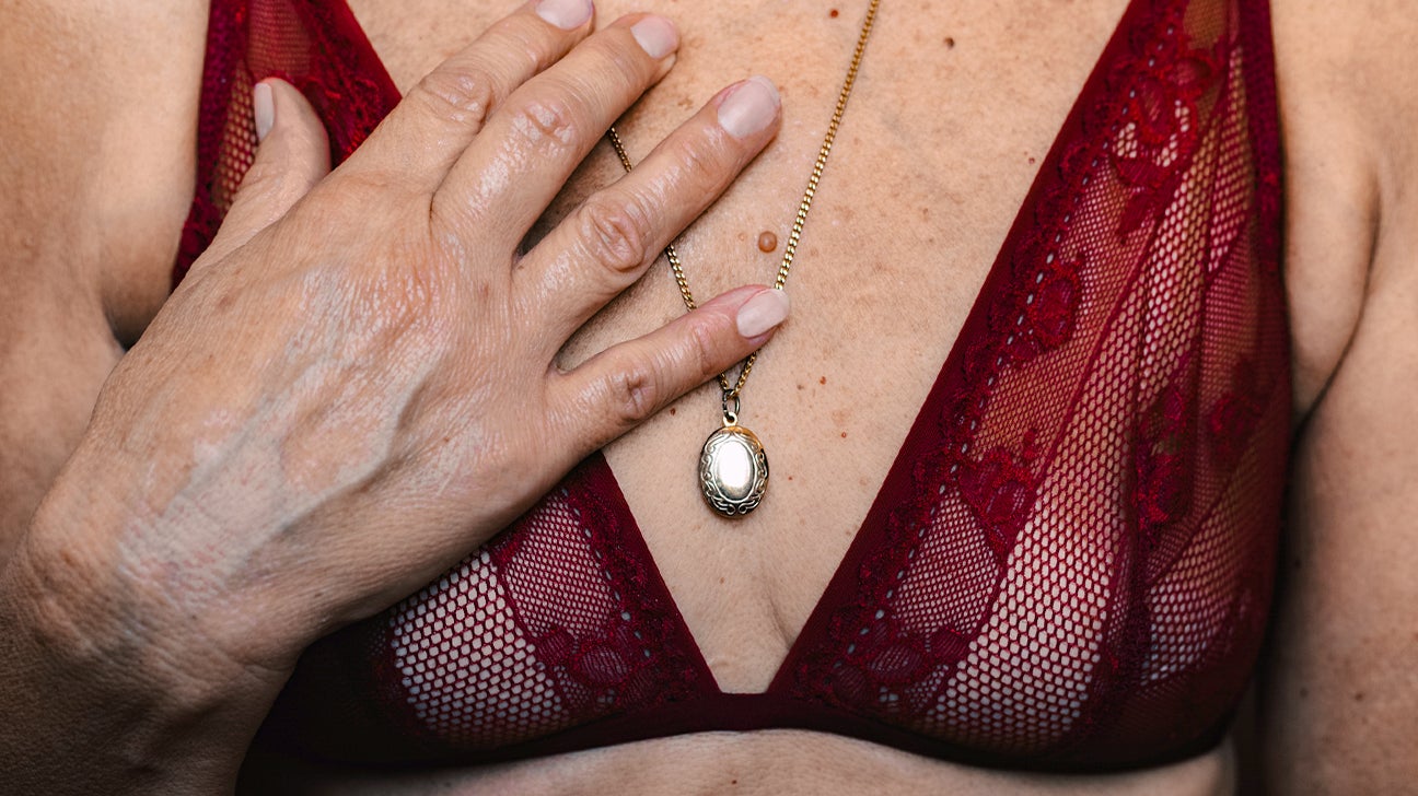
- 💕Breast Cancer💕 Did you know at the age of 38 I felt a lump while taking a shower? I felt something and I did something. I booked
- Wholesale sexy boob size - Offering Lingerie For The Curvy Lady