Red and white blood cells in clot, SEM - Stock Image - C045/8688 - Science Photo Library

By A Mystery Man Writer
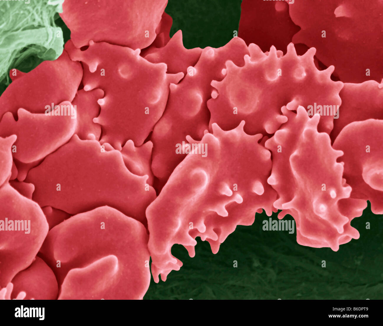
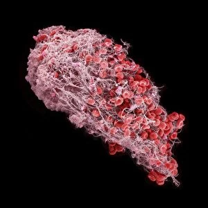
Red blood cells (erythrocytes) and a single white blood cell (leucocyte or leukocyte) in a fibrin mesh, coloured scanning electron micrograph (SEM). Formation of a blood clot with many erythrocytes (red) and a single leukocyte (white/blue) becoming entangled in a fibrin mesh (light brown). ANNE WESTON, FRANCIS CRICK INSTITUTE/SCIENCE PHOTO LIBRARY

Human Red Blood Cells Sciencephotography.com

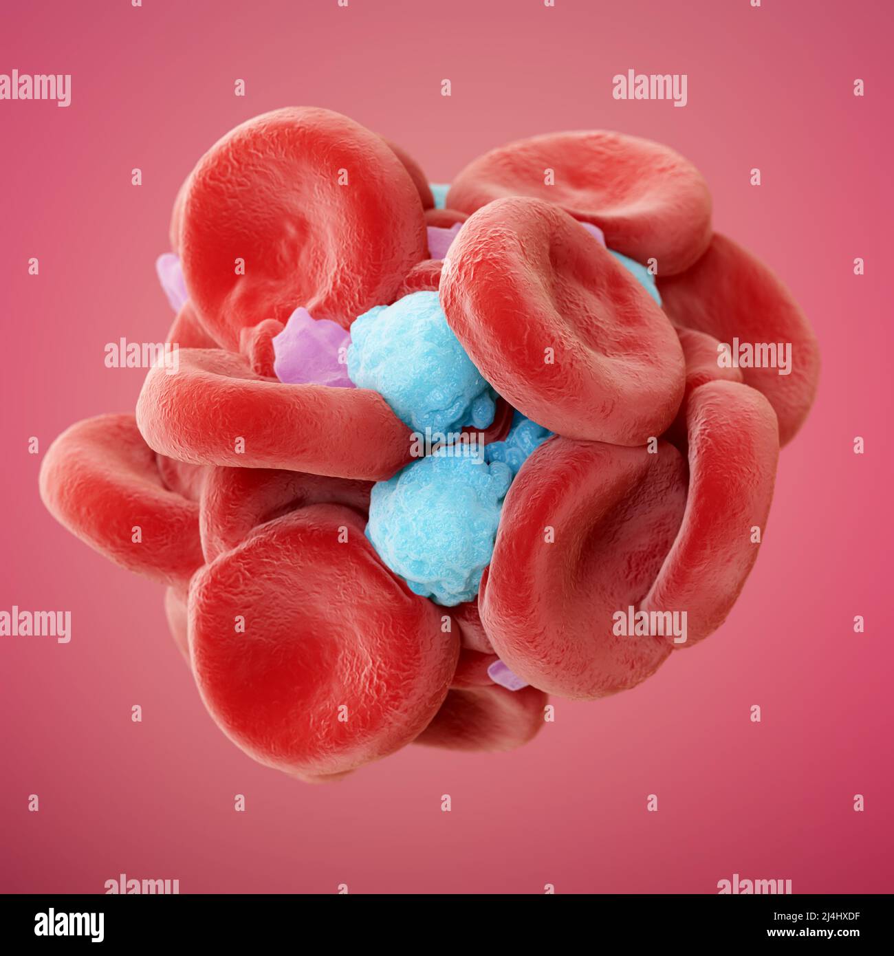
Science Photo Library - Illustration of a blood clot. Depicted

Science Photo Library - Leukaemia blood cells. Composition

Red blood cells, white blood cells and platelets, SEM - Stock
Blood clot. Coloured Scanning Electron Micrograph (SEM) of a blood
Fibrin red white blood hi-res stock photography and images - Alamy

Red and white blood cells in clot, SEM - Stock Image - C045/8688

Red and white blood cells in clot, SEM - Stock Image - C045/8688
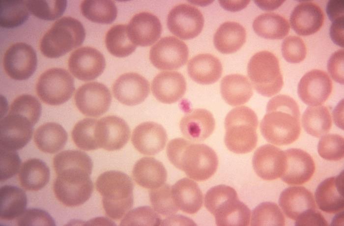
Details - Public Health Image Library(PHIL)
Blood clot, coloured scanning electron micrograph (SEM). Red blood cells (erythrocytes) are trapped within a fibrin protein mesh (beige). The fibrin
Prints of Blood clot, SEM C016 / 9745

31,828 Clot Royalty-Free Photos and Stock Images

Blood clot, SEM - Stock Image - C056/3890 - Science Photo Library

Blood Clot, Sem #7 by Steve Gschmeissner
- LEDs on lettuce: White light versus red + blue light - Produce Grower
_fmt.png)
- Lynch Sign 5 ft. x 3 ft. Red on White Vinyl Under New Management

- Grunge Red Under Review Word Round Rubber Seal Stamp On White

- Boys' UA Leadoff Mid RM Jr. Baseball Cleats | Under Armour
- Long amazing hair with platinum blonde above and red underneath!

- Women's Underwear Tube Top Off Shoulder Bra Lace Bralette Sleeveless Wedding Dress Female Underwear

- Hue Studio : Leggings for Women : Target
- Gameday Couture San Francisco 49ers Women's White Top Recruit

- Nvgtn Leggings - Canada

- Leonisa Leggings Women's Shapewear: Bodysuits, Waist Trainers & More! - Macy's