Histology, microscopy, anatomy and disease: Week 3: 2.1
By A Mystery Man Writer
Histology, microscopy, anatomy and disease: Week 3: 1.1

Shielding islets with human amniotic epithelial cells enhances islet engraftment and revascularization in a murine diabetes model - American Journal of Transplantation

Clinical Validation of Stimulated Raman Histology for Rapid Intraoperative Diagnosis of Central Nervous System Tumors - Modern Pathology

Anatomy, Histology, and Normal Imaging of the Endometrium
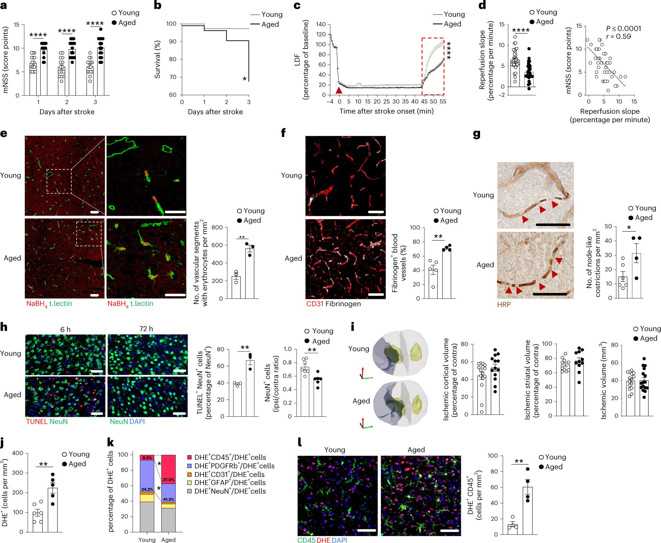
Age-induced alterations of granulopoiesis generate atypical neutrophils that aggravate stroke pathology
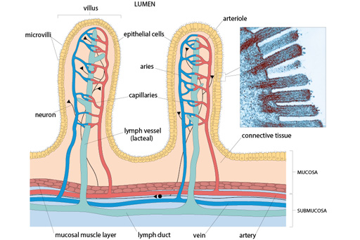
Histology, microscopy, anatomy and disease: Week 2: Figure 3 Cross-section of the gut showing the arrangement of the different tissue elements at the level of the ileum (small intestine).

Free Course: Histology, microscopy, anatomy and disease from The Open University
Normal Human Histology, Large Set, Part II. - English Slides - 1004235 - W13410 - LIEDER - 72000_EN - English - 3B Scientific

Quantitative 3D microscopy highlights altered von Willebrand factor α‐granule storage in patients with von Willebrand disease with distinct pathogenic mechanisms - Research and Practice in Thrombosis and Haemostasis

2.4.3 Skin Pathology I Flashcards by Priyanka Bhandari
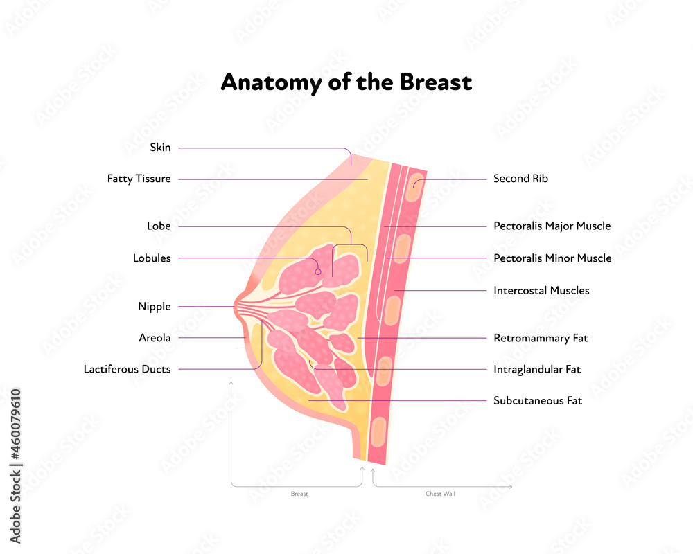
- Breast anatomy, artwork - Stock Image - C008/4452 - Science Photo Library
- Schematic diagram showing the anatomy of the breast.
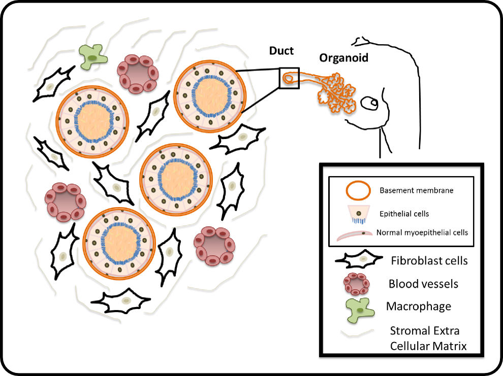
- Breast Cancer — Cellular and Molecular Biomechanics Laboratory
- The BMJ on X: Diagram showing common sites and types of breast

- Human breast anatomy diagram. Vector flat medical illustration

- Women Sexy Bra 32G Bras Women Fabric Nipple Cover Coconut Shampoo

- KILLSTAR Neo Noir Leggings

- Dacada2005 12 panties colors STV size M-L-XL-XXL-3XL-4XL seamless Lycra women underwear high

- fuel tank level lid sensor lock

- ZYIA Active All Star One More Rep NO PadS Longling High Neck Sports Bra Small S – St. John's Institute (Hua Ming)
