Ultrasound imaging
By A Mystery Man Writer
Ultrasound imaging - Download as a PDF or view online for free
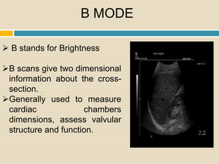
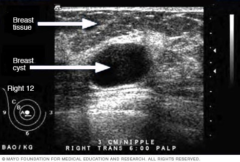
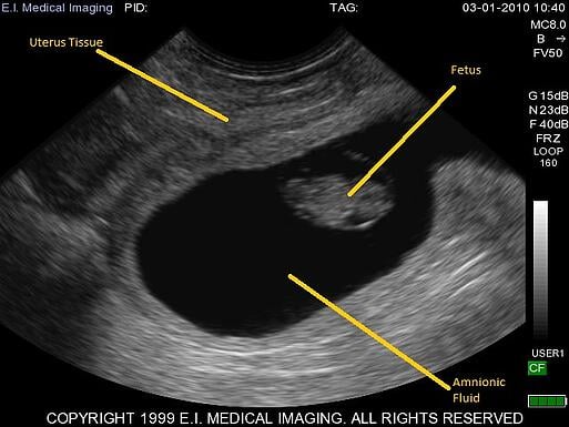
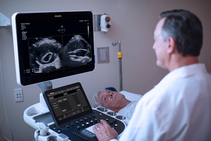
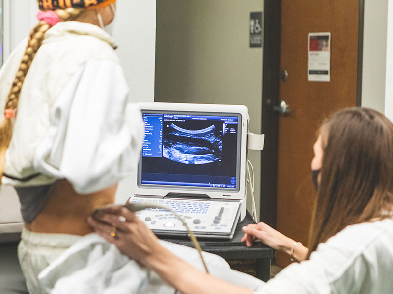
Ultrasound uses high frequency sound waves to visualize internal structures. It works by transmitting sound waves into the body using a transducer probe, which detects the echoes as they bounce off tissues and organs. The echoes are processed to form images on the ultrasound machine screen in real-time. Common applications include obstetrics, cardiology, and urology. The Philips HD11 is an ultrasound system with curvilinear, linear, and phased array probes for different exams. It provides grey scale, Doppler, and color imaging modes. Ultrasound has benefits of being non-invasive, portable, and having no radiation, but has limitations of being operator dependent and unable to penetrate bone.
Ultrasound - Mayo Clinic
Ultrasound Basics: How to Read an Ultrasound Image
Ultrasound imaging (A) when the transducer is placed in the horizontal
Ultrasound rises to the challenges of 21st century imaging - Blog

Rehabilitative Ultrasound Imaging (RUSI)

Special probes improve ultrasound imaging in obese patients
Rehabilitative Ultrasound Imaging (RUSI)
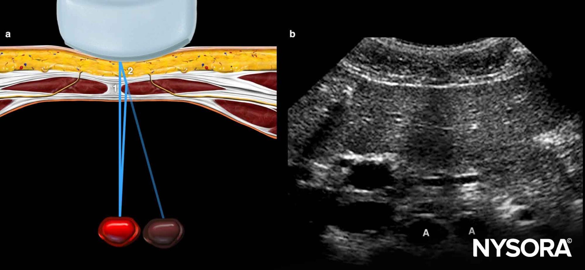
Ultrasound image artifacts explained - NYSORA
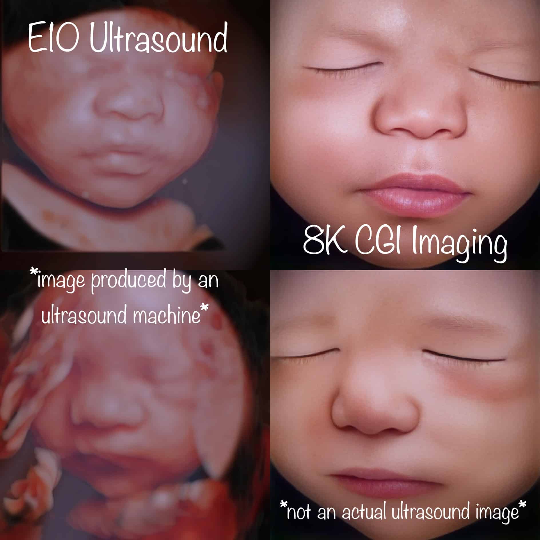
Unraveling the Mystery of Ultrasound Imaging: 2D, 4D, 5D, 8K, and HD Edit Explained - Enlightened 4D Imaging

4 Ways To Improve Your Ultrasound Imaging

Understanding Ultrasound: Common Uses and Benefits
- A) A B-mode ultrasound image of a bladder in a transverse section

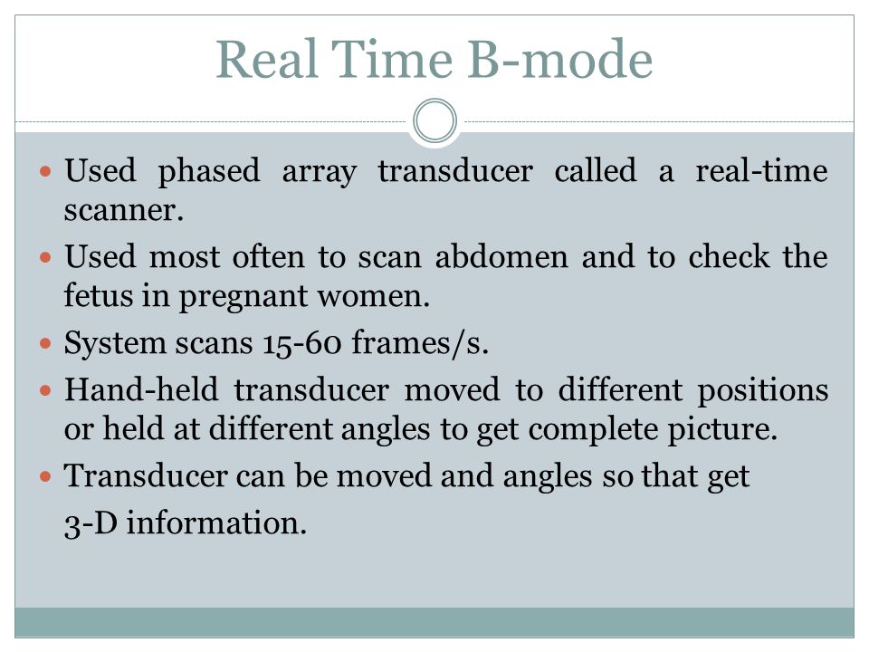
- Modes Ultrasound A-mode- amplitude mode. B-mode- brightness mode. - ppt video online download
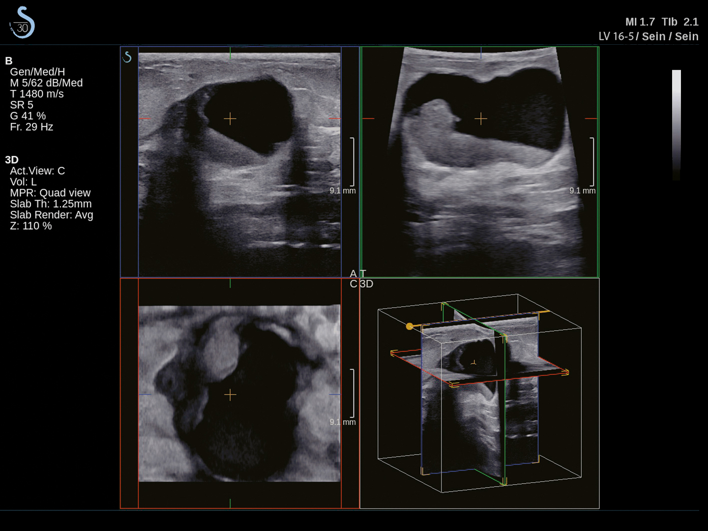
- BREAST / Aixplorer MACH / Home - Supersonic Imagine
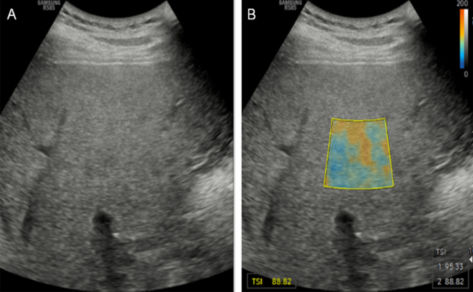
- Quantitative ultrasound imaging of soft biological tissues: a primer for radiologists and medical physicists, Insights into Imaging
- TE Air - Mindray Wireless Handheld Ultrasound System - Mindray

/product/34/743534/1.jpg?8189)


:strip_icc()/Neutral-dressing-room-101960176_3Bxm9OtqqdH931z2spK7M_-77dd8c53e058437db7a2b4581956f62b.jpeg)