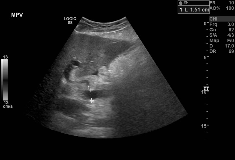
Figure, B-Mode ultrasound showing main portal] - StatPearls
By A Mystery Man Writer
B-Mode ultrasound showing main portal vein diameter of 15.1 millimeters. This is an indirect finding of portal hypertension. Contributed by Brian Covello, MD

Operator Evaluation of Ultrasound Fusion Imaging Usefulness in the Percutaneous Ablation of Hepatic Malignancies: A Prospective Study - ScienceDirect
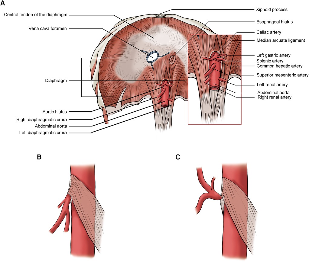
Frontiers Ultrasound characteristics of abdominal vascular compression syndromes
Salivary gland ultrasound in primary Sjögren's syndrome
B mode ultrasound image (A) shows the hypoechoic tract (arrow) which is

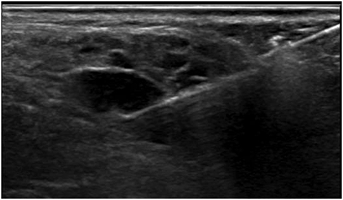
Sonography of a Typical Parathyroid Adenoma: Solitary Parathyroids as Seen on Ultrasound
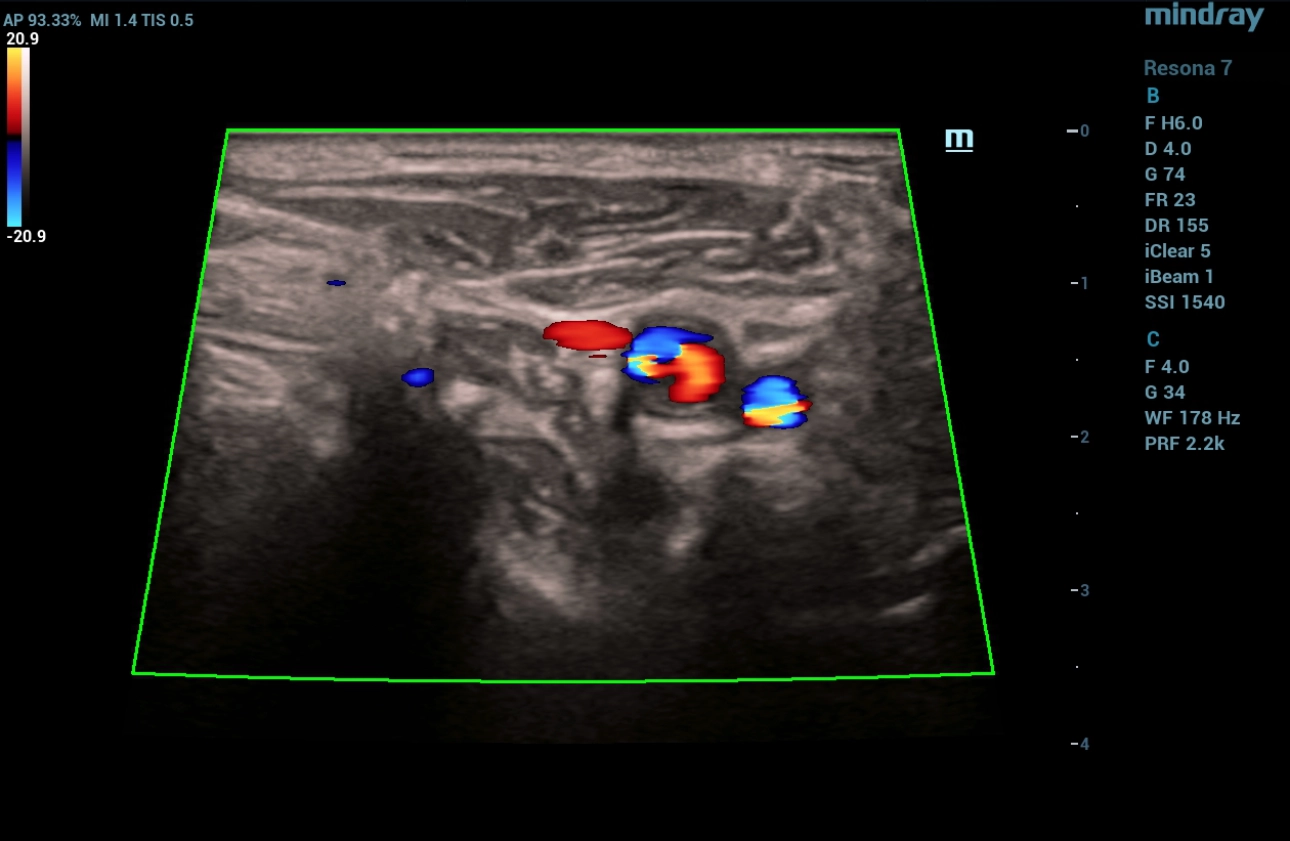
Ultrasound Journal 23 - Postoperative Ultrasound: A Case Study in Cardiovascular Pathology - Mindray

Gallbladder sludge, Radiology Case
Gastroenterology Insights, Free Full-Text

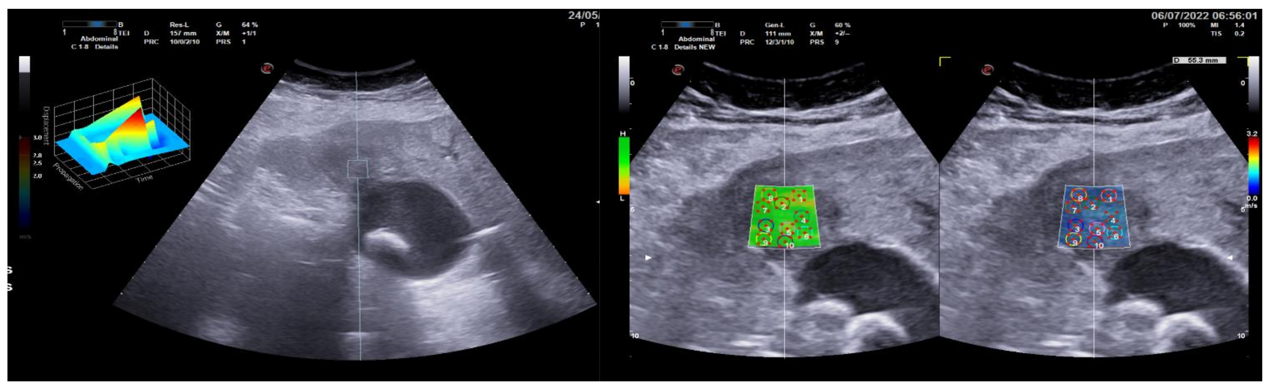
Tethered spinal cord tension assessed via ultrasound elastography in computational and intraoperative human studies

Pathogens, Free Full-Text
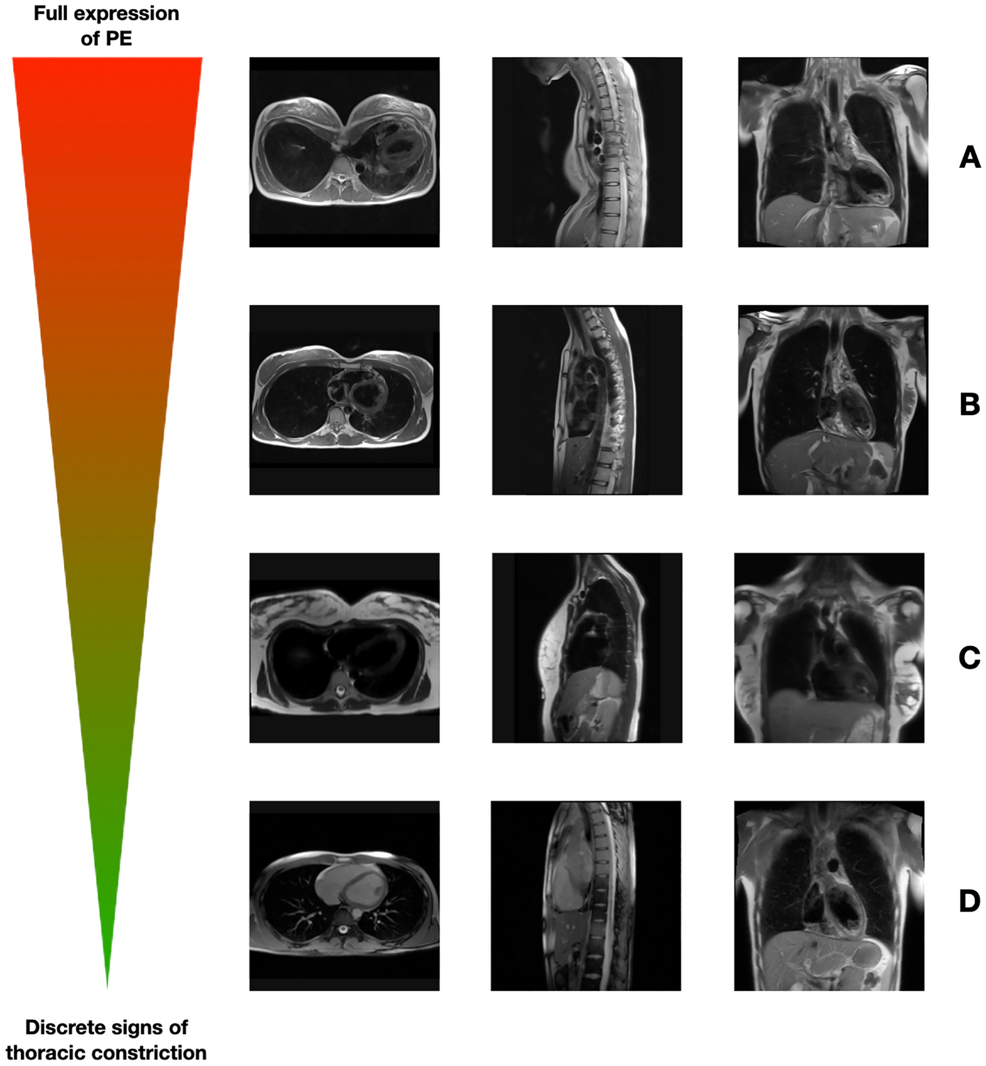
Description of a new clinical syndrome: thoracic constriction without evidence of the typical funnel-shaped depression—the invisible pectus excavatum

Point of Care Abdominal Ultrasound - ScienceDirect

Ross Hauser, MD Reviews Cervical Spine Instability and Potential Effects on Brain Physiology
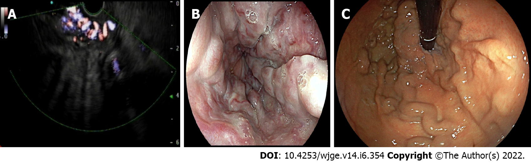
Role of endoscopic ultrasound in vascular interventions: Where are we now?

Point of Care Abdominal Ultrasound - ScienceDirect
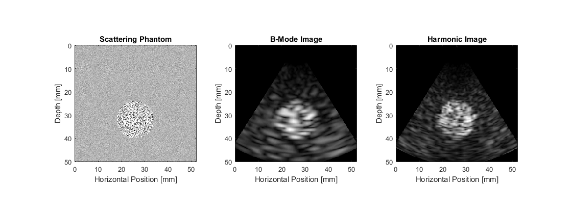
- k-Wave MATLAB Toolbox
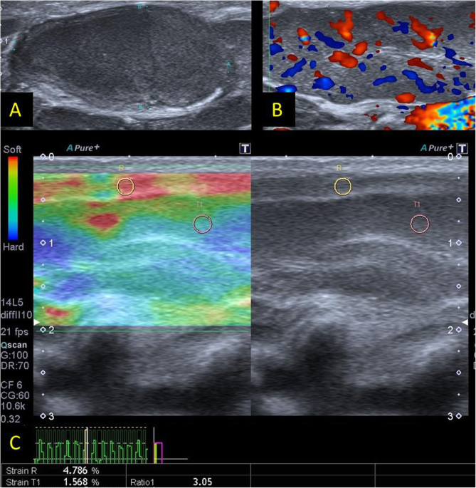
- B-mode ultrasound, color Doppler, and sonoelastography in differentiation between benign and malignant cervical lymph nodes with special emphasis on sonoelastography, Egyptian Journal of Radiology and Nuclear Medicine
- Ultrasound Modes, A, B and M Mode, Ultrasound Physics

- Non-contrast power Doppler ultrasound imaging for early assessment
- Radiogenomic Analysis of Breast Cancer by Using B-Mode and

- Fitness Fashion: Understanding Consumer Preferences in the Activewear Market, by Soniyakale, Feb, 2024

- ASOS DESIGN skinny fit t-shirt bodysuit in white
- Women Buttock Underwear Briefs Knickers Bum Lift Shaper Enhancer Pants Push Up

- KIWI RATA High Waist Scrunched Butt Leggings for Women Compression Fitness Yoga Pants Butt Lift Activewear Tights

- Avalanche Womens Drawstring Waistband Legging Fitted Jogger

