Figure 6 from Femoral Hernia: A Review of the Clinical Anatomy and

By A Mystery Man Writer
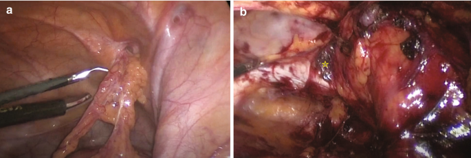
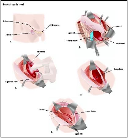
Figure 6. Femoral hernia repair in clean operation. (a) The narrow side of the mesh is sutured to Cooper’s ligament; (b) The mesh is sutured to the iliopubic tract or shelving portion of the inguinal ligament; (c) The posterior wall of the inguinal canal is reinforced, as in Lichtenstein’s repair. - "Femoral Hernia: A Review of the Clinical Anatomy and Surgical Treatment"
Femoral Hernia
Femoral Hernia - Risk Factors - Clinical Features - Management - TeachMeSurgery

Richter Hernia: Surgical Anatomy and Technique of Repair

Atypical femoral hernia called Laugier's hernia: clinical features, radiological findings, and management at a single center
Abdominal Hernia - Epigastric - Spigelian - Obturator - TeachMeSurgery
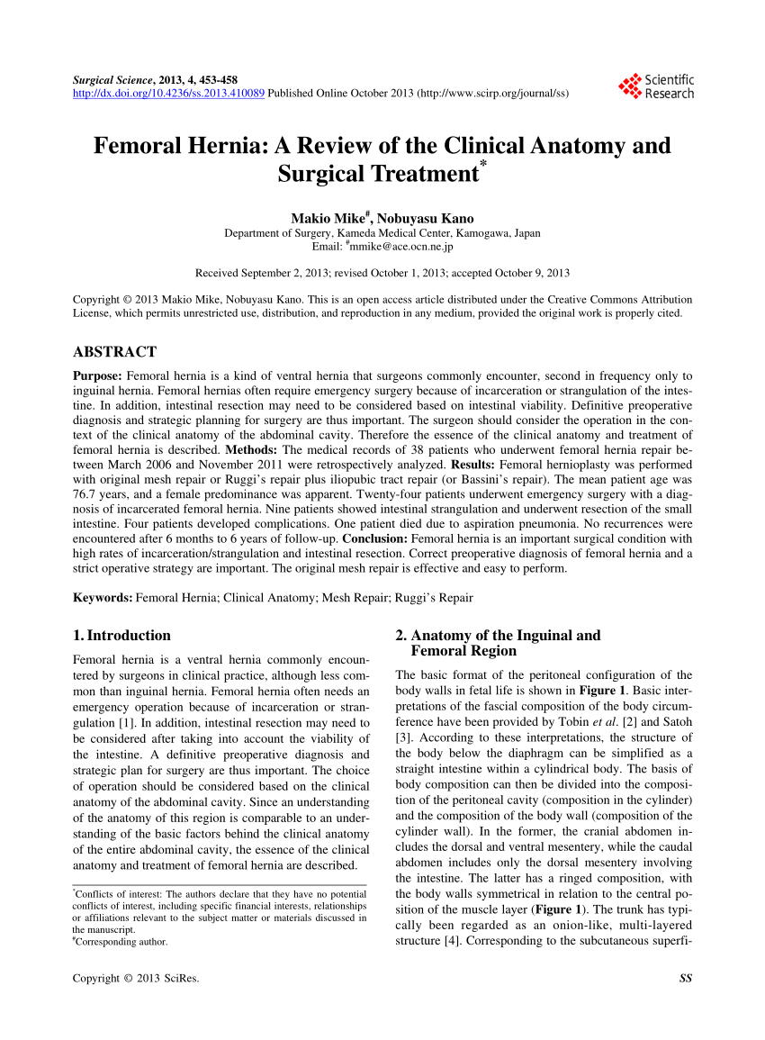
PDF) Femoral Hernia: A Review of the Clinical Anatomy and Surgical Treatment

Femoral Hernia - A Review of Clinical Anatomy
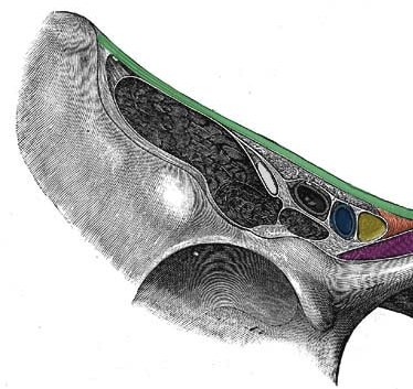
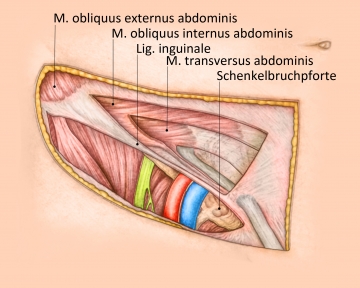
Myopectineal orifice. The oval-shaped myopectineal orifice (green

Figure 4 from Femoral Hernia: A Review of the Clinical Anatomy and Surgical Treatment

Adult groin hernias - ScienceDirect
Anatomy of the inguinal and femoral regions. (A) Transversalis fascia

AIS Channel · Hernia Surgery
- Anatomy - Femoral hernia repair – TIPP technique
- Femoral Hernia, Mr. Himaz Marzook

- Femoral Hernia Repair - procedure, recovery, blood, pain, complications, adults, time, infection
- Keyhole Surgery for Inguinal & Femoral Hernia in Adult Melbourne
- Open femoral hernia repair reveals a hernia sac containing a necrotic


:upscale()/2017/08/11/820/n/1922564/867e0465ac7e8960_ZIPPER_JEAN_CELTIC_2.jpg)