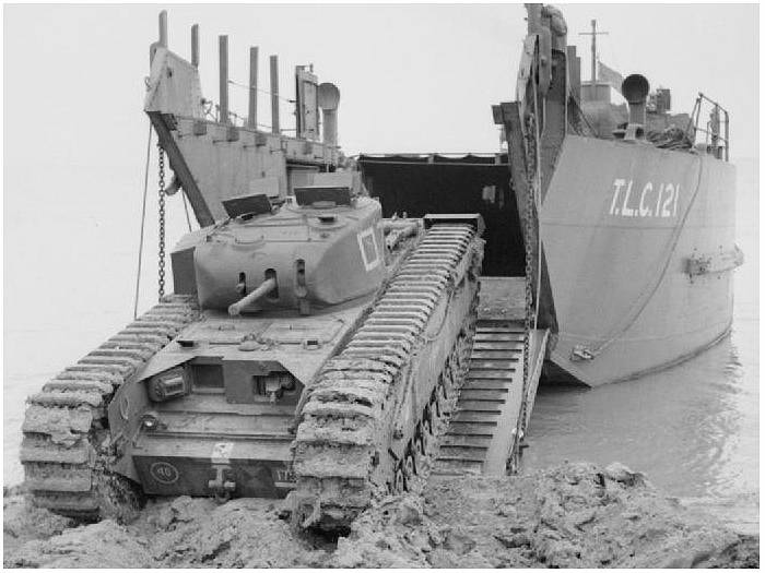
Comparison of MRI slices at the mid calf showing the anatomy with an IP

By A Mystery Man Writer
Applications of radiological imaging in endocrine disease (5C) - Endocrine Pathology

Gulf View Medical Centre - Did you know MRI's are commonly used to examine the brain, spine, joints, abdomen, and pelvis? Below are some imaging facts and information to consider: • An

Macroscopy of tissue-engineered bi-and tricuspid venous valves.
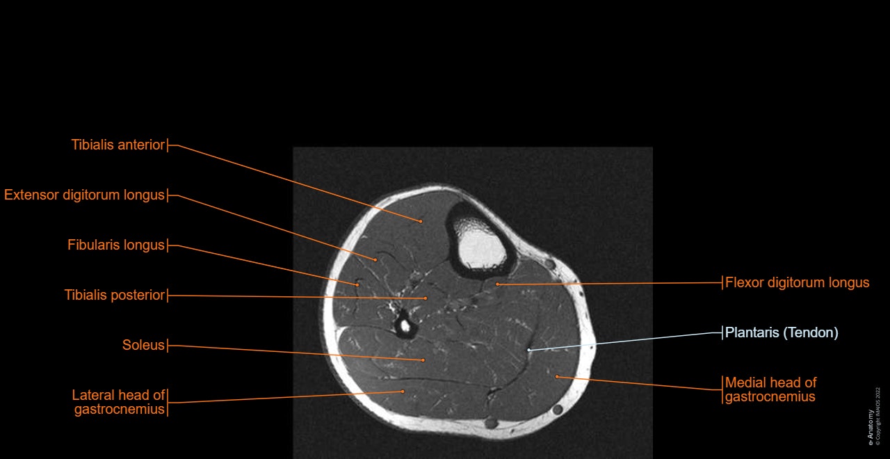
Lower limb: MRI anatomical atlas
MIDA: A Multimodal Imaging-Based Detailed Anatomical Model of the Human Head and Neck

Jean-Patrick BENIGNI, Medical Doctor, Polytech Paris-UPMC, Paris, polytech paris, Département de pédagogie

Hippocampal volumetric variations in the normal human brain by magnetic resonance imaging (MRI)

PDF) Relationship between medical compression and intramuscular pressure as an explanation of a compression paradox

The occurrence and prevalence of pathological reflux in segments

André CORNU-THÉNARD, Vascular Medical Doctor, Research in Compressiontherapy and Treatment of Elephantiasis by Reduction, Cornu-Thenard MD, FACPh -, Doctor André Cornu-Thenard; France, Paris, 75011 Faidherbe Street 2

Jean-François UHL, Unesco Chair of digital anatomy, MD, FacPh, Paris Descartes, CPSC, Paris, Paris 5, UFR biological (Saints-Pères)

Comparison of high‐field and low‐field magnetic resonance images of cadaver limbs of horses - Murray - 2009 - Veterinary Record - Wiley Online Library

Comparison between right and left limbs.
- Men's Half Calf Socks – Bombas

- Polynesian half calf sleeve done my Tripps Kahai at Ascension tattoo in Orlando, Florida : r/tattoos
- WOLF CREEK DAIRY - Got our first half Holstein half Angus calf yesterday, does that make then Holguses or Angsteins?
- Sedley's Half Calf

- Hughez White Platform Mid-Calf Boots