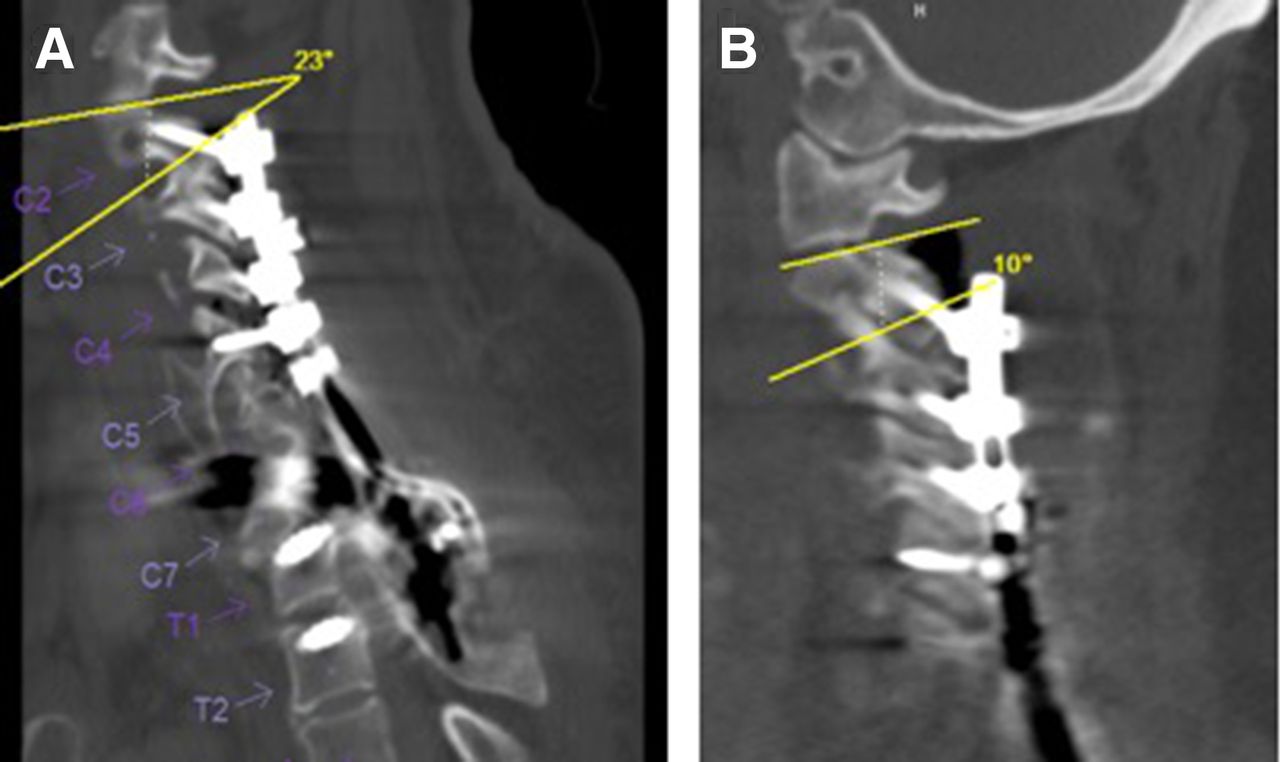
CT image of C2–3 congenital fusion. A Vertebral body fusion and lamina

By A Mystery Man Writer

Malformed vertebrae: a clinical and imaging review, Insights into Imaging

Anatomical analysis of the C2 pedicle in patients with basilar invagination
The Spine: Congenital and Developmental Conditions

Figure 5 from Congenital fusion of cervical vertebrae clinical significance

CT image of C2–3 congenital fusion. A Vertebral body fusion and lamina

Anatomical analysis of the C2 pedicle in patients with basilar invagination
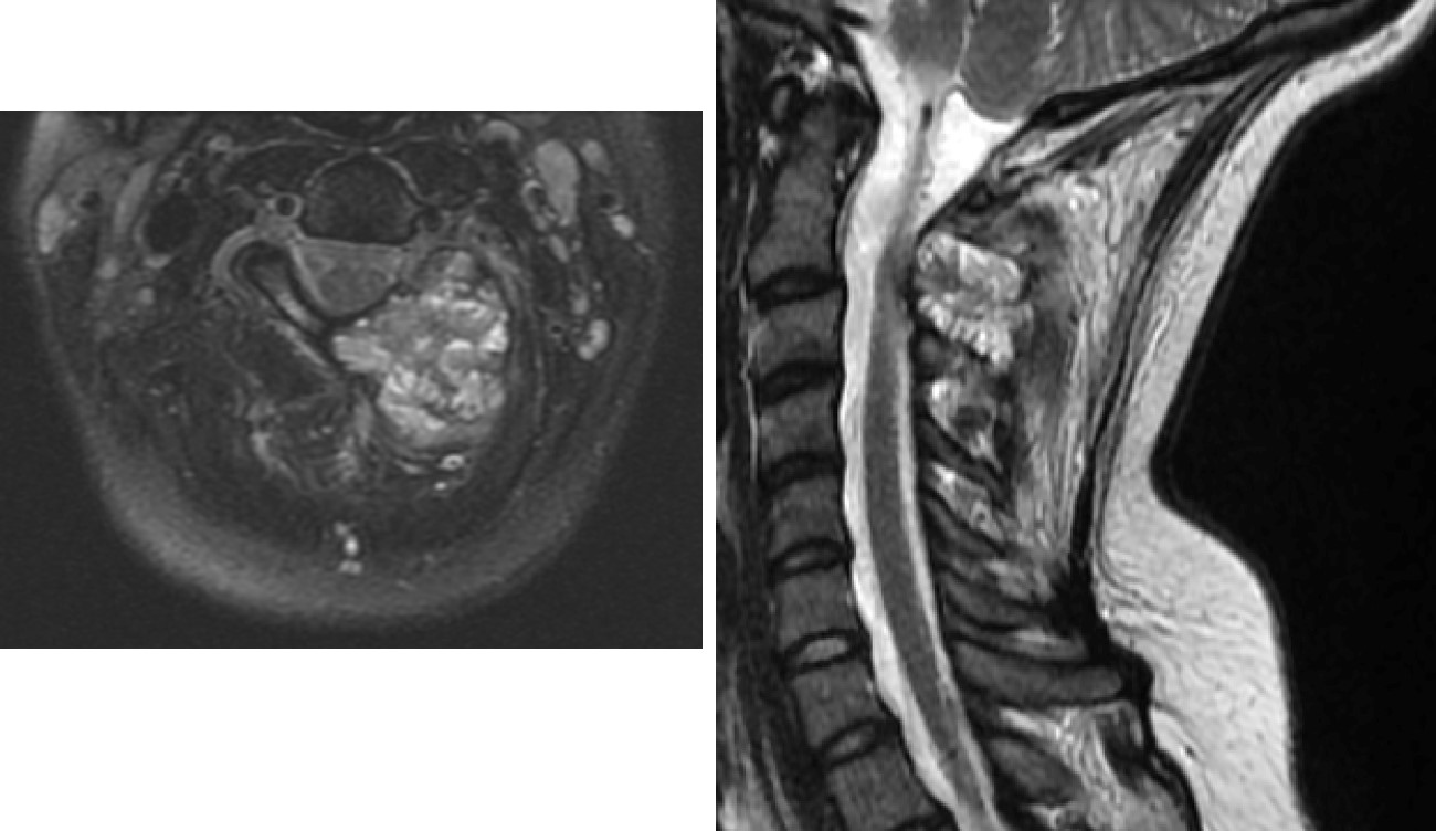
Anterior–Posterior Surgical Treatment of a C2 Aneurysmal Bone Cyst
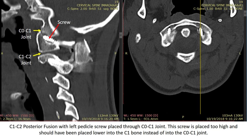
C1-C2 Fusion - Complications Are Common

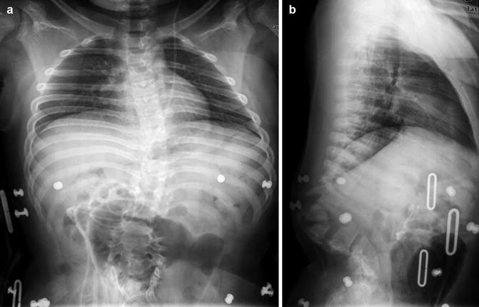
Congenital C2-C3 vertebral fusion, Radiology Case
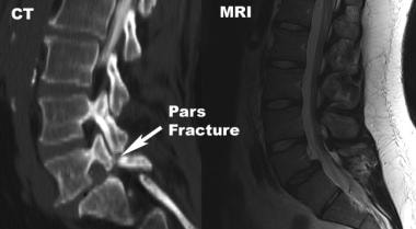
Spinal Instability and Spinal Fusion Surgery Workup: Laboratory Studies, Imaging Studies, Other Tests

Craniocervical instability in patients with Ehlers-Danlos syndromes: outcomes analysis following occipito-cervical fusion
C1-C2 Facet Joint Penetration by C2 Pedicle Screws: Influence of Local Anatomy, Bone Mineral Density, and Screw Length