A) A brightness mode (b-mode) image of the lateral abdominal wall.

By A Mystery Man Writer
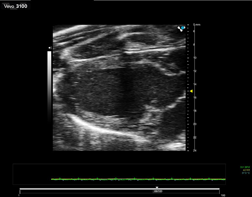
Download scientific diagram | (A) A brightness mode (b-mode) image of the lateral abdominal wall. Abbreviations: EO, external oblique; IO, internal oblique; TrA, transversus abdominis. (B) A split-screen image with b-mode on the left and motion mode (m-mode) on the right. The m-mode image represents the information from the dotted line on the b-mode image displayed over time (x-axis). Static structures produce straight interfaces while structures that change in thickness or depth (in this case the TrA) create curved interfaces. The increase in depth of the TrA correlates to a contraction. Reproduced with permission Whittaker 2007. 142 from publication: Rehabilitative Ultrasound Imaging: Understanding the Technology and Its Applications | The use of ultrasound imaging by physical therapists is growing in popularity. This commentary has 2 aims. The first is to introduce the concept of rehabilitative ultrasound imaging (RUSI), provide a definition of the scope of this emerging tool in regard to the physical | Rehabilitation, Ultrasonography and Ultrasound Imaging | ResearchGate, the professional network for scientists.

A) A brightness mode (b-mode) image of the lateral abdominal wall.

A) A brightness mode (b-mode) image of the lateral abdominal wall.

A) A brightness mode (b-mode) image of the lateral abdominal wall.
Ultrasonic Imaging: Physics and Mechanism
Basics of Ultrasound: Pitfalls and Limitations - NYSORA

Modes Ultrasound A-mode- amplitude mode. B-mode- brightness mode. - ppt video online download
Jackie WHITTAKER, Professor (Associate), BScPT, PhD
Physics and Instrumentation in Doppler and B-mode Ultrasonography
Frontiers Preclinical Ultrasound Imaging—A Review of Techniques and Imaging Applications
.jpg)
The A, B, M's – Ultrasound Modes Explained

A) A brightness mode (b-mode) image of the lateral abdominal wall.

Bridging repair of the abdominal wall in a rat experimental model. Comparison between uncoated and polyethylene oxide-coated equine pericardium meshes

A) A brightness mode (b-mode) image of the lateral abdominal wall.

Brightness mode ultrasound (B-mode): grayscale ultrasound showing fiber